[!NOTE]
What you are doing: Mapping the vermillion border and identifying mucosal tissue transitions.
Why you are doing it: Tattooing outside the vermillion into perioral skin heals as an unnatural clown-like outline.
How you know it is right: Pigment is strictly confined to the vermillion border and modified vermillion zone.
Lip tissue is fundamentally different from facial skin. It lacks hair follicles, sweat glands, and has an ultra-thin stratum corneum with extensive underlying vascularity:
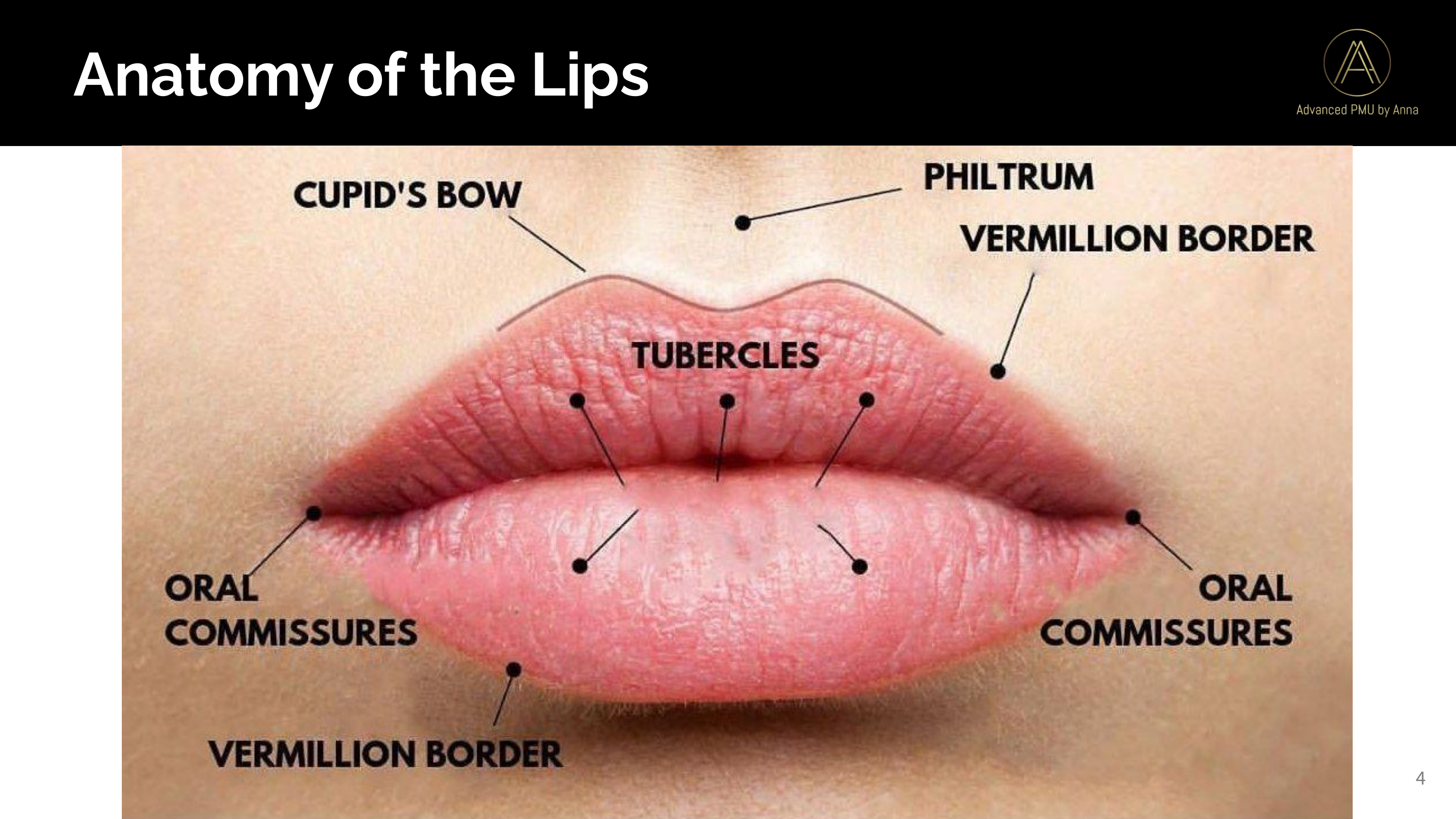
Key Lip Anatomical Structures & Clinical Boundaries
| Structure | Anatomical Description | Histological Characteristic | Clinical Tattooing Boundary Rule |
|---|---|---|---|
| Vermillion Border | Demarcated perimeter between facial skin and red lip | Modified stratified squamous epithelium | The absolute outer boundary. Never tattoo past this into pore-bearing skin. |
| Cupid's Bow | Central dual peaks and trough of the upper lip | Peak convergence of philtral columns | Follow natural anatomical curvature; never flatten or artificially over-draw. |
| The Wet-Dry Line | Inner boundary where dry vermillion meets wet oral mucosa | Non-keratinized, mucus-secreting epithelium | The strict inner stop boundary. Tattooing past this causes pigment sloughing. |
| Oral Commissures | The left and right corners of the mouth | Moist, highly mobile mucosal folds | Stop shading 1mm to 2mm before the true corner to prevent pooling and tearing. |
| Philtral Columns | Two vertical ridges running from nasal septum to Cupid peaks | Fibrous dermis with elevated ridges | Used as vertical symmetry reference points during pre-draw mapping. |
| Fordyce Spots | Tiny 1mm to 2mm yellow or white granules on vermillion | Benign, ectopic sebaceous glands | Normal anatomical variation. Never needle aggressively over dense clusters. |
Histology & Vascularity: Why Lips Swell and Bruise
The red coloration of the vermillion is caused by extensive capillary loops in the dermal papillae showing through thin, translucent epithelial layers. High needle trauma or excessive machine voltage ruptures these superficial capillaries, resulting in rapid hematoma (bruising) and intense edema (swelling). Maintain light, airbrush-like pressure with a 45 to 60 degree machine angle.
The Vermillion Boundary Rule: Preventing the "Clown Lip" Trap
One of the most catastrophic mistakes in lip permanent makeup is attempting to enlarge thin lips by tattooing outside the vermillion border into perioral facial skin:
- Pore and Follicle Mismatch: Perioral facial skin contains visible sebaceous pores, hair follicles, and a thick stratum corneum. In contrast, the vermillion has zero hair follicles and an ultra-thin, translucent surface.
- Healed Optical Disconnect: When pigment heals in facial skin, it reflects light completely differently than pigment in mucosal tissue. While the color on the vermillion heals as a soft, translucent watercolor tint, the pigment on the facial skin heals as an opaque, muddy, unnatural ring around the mouth.
- Long-Term Aging: As the client speaks, smiles, and ages, dynamic facial muscles move independently of the lip border. Pigment placed on the skin will emphasize perioral wrinkling and look like smeared, mismatched lipstick that never washes off.
Managing Fordyce Spots with Care
Fordyce spots are benign, ectopic sebaceous glands that appear as small, isolated or clustered white-to-yellowish granules beneath the mucosal surface. They are present in over 70% of adult lips and represent completely normal anatomy.
- The Over-Needling Risk: Fordyce spots contain sebum and lack normal dermal papillae. If an artist attempts to "cover" or tattoo over dense Fordyce clusters with heavy pressure or repeated passes, the traumatized glands become inflamed, reject pigment, and heal as swollen, raised white bumps.
- Clinical Protocol: Glide lightly across areas with Fordyce spots using a 3RS or soft 1RL at low voltage. Never stop to pack pigment directly into a cluster. Educate the client beforehand that permanent makeup enhances lip tone, but cannot erase underlying sebaceous gland structures.
Visual Reference Summary